High precision keratometry & topography

For fewer toric surprises

Dual zone keratometry

Highly precise Ks

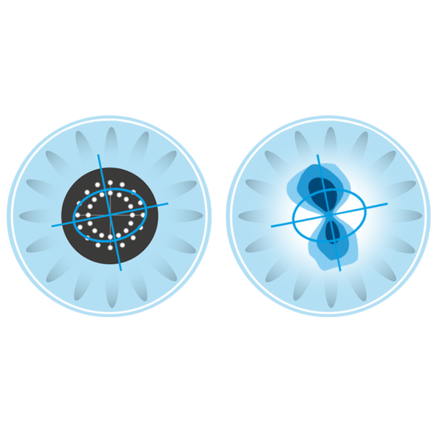
Lenstar 900’s gold standard dual zone keratometry provides precise measurements of astigmatism and the axis position, making it the biometer of choice for toric IOLs. Precision is further enhanced due to its closely spaced 32 measurement point pattern that relies on real, measured data instead of software data interpolation of just a few points.
T-Cone topography

Double-check the axis

For increased confidence in toric procedures, the optional T-Cone toric platform adds true 11-ring Placido topography of the central 6 mm optical zone to Lenstar 900’s offering. K-readings optioned with the T-Cone are derived using the same algorithms as with the standard Lenstar 900 without T-Cone and are therefore interchangeable. Equipped with this additional information, surgeons can double check axis position and astigmatism for optimal planning of toric IOLs.

Full information

Identification of irregular astigmatism

Irregular astigmatism is a counterindication to the use of toric IOLs. To identify it with ease, the optional T-Cone platform offers axial, tangential and elevation maps as well as the color images of the Placido rings. This allows full assessment of the suitability of a patient as a toric candidate and supports patient education.

T-Cone Add-on

Placido topography

The optional T-Cone platform is a cost-effective solution to provide topography information in practices that lack a topographer. It can be mounted at any time.

Keratometry overview

Detailed data review and quality management

The keratometry overview screen provides a concise and comprehensive overview of all relevant data. This enables an efficient review of the image quality of the keratometry measurement points related to the patient's tear film stability. Using this information allows a user to make an informed decision about which specific datapoints to include and which ones to exclude from the final examination calculation.
Topography overview

Topography data review and quality management

The topography overview screen provides a concise and comprehensive overview of captured topography information and includes axial and tangential curvature as well as elevation maps and images of Placido ring reflections for each individual measurement taken. This allows an efficient review of data and image quality and supports detection of artefacts such as a torn tear film break up or obstructing eye lashes. In addition, users can easily include or exclude individual measurements to obtain optimum topography results.
Axial curvature map

Assessment of regularity of the astigmatism

The axial curvature map, sometimes also referred to as the sagittal map or power map, is the most commonly used topography representation. It allows to gain a good overall perspective on the regularity of the astigmatism and thus enables rapid decision making about a patient’s suitability for a toric IOL.
Tangential curvature map

Identification of local irregularities

The tangential curvature map, sometimes also referred to as the local curvature map or instantaneous rate of curvature, enables detection of more subtle, localized corneal curvature irregularities. This is especially useful for early detection of corneal pathology such as Keratoconus.
Elevation map

Detection of local elevations or depressions

The elevation map provides insights into how much the cornea deviates from a best-fit sphere of the cornea. This allows identification of small elevations or depressions that may be caused by pathologies or have been induced during surgery. This maps has proven to be an excellent tool for post-refractive documentation of surgery.
arrow-right
arrow-left
Keratometry overview
Topography overview
Axial curvature map
Tangential curvature map
Elevation map