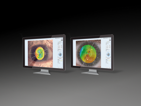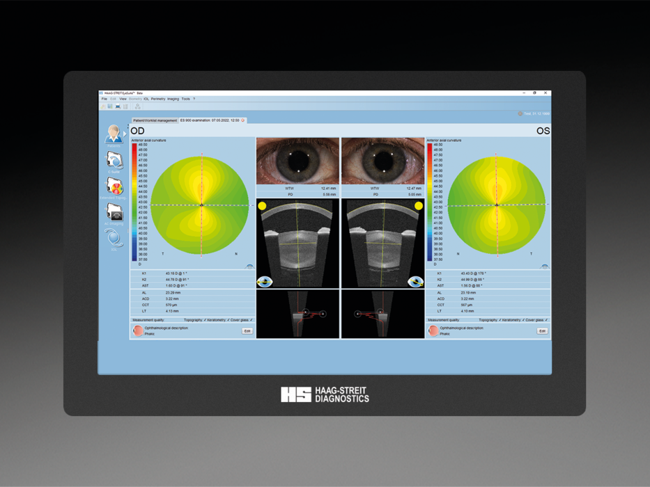
High-precision data for cataract and refractive surgery since 2008
Biometry
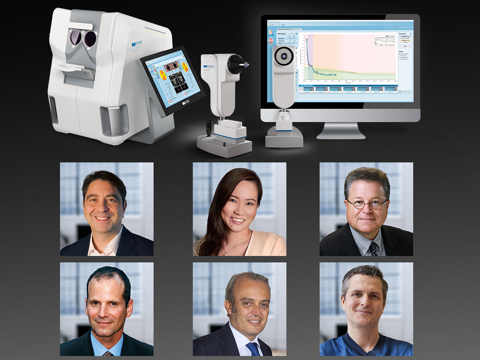
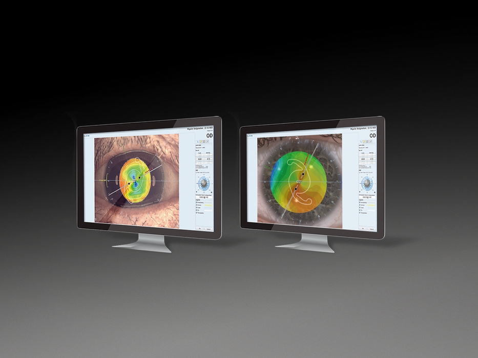
Haag-Streit optical biometers support the daily quest of the cataract and refractive surgeon and their ophthalmic technicians to provide excellent refractive results to the patient. Our Lenstar and Eyestar biometers deliver precise measurements, intuitive data representation, and latest-generation planning & analysis tools to support reliable diagnosis and surgery. Quick measurement acquisition, automation support, and ergonomic user interfaces ensure that the process of diagnostic data acquisition is pleasant for surgeon and patient alike.
Compare our products
| Eyestar 900 | Lenstar 900 | |
|---|---|---|
| Applications | ||
| Cataract surgery | ● | ● |
| Refractive surgery | ● | ● |
| Anterior chamber analysis | ● | − |
| IOL calculation | ● | ● |
| Premium IOL calculation | ● | ○ |
| Myopia management | ○ | ○ |
| Biometry data | ||
| Biometry (AL, ACD, LT, CCT) | ● | ● |
| Keratometry (Dual zone anterior) | ● | ● |
| Anterior corneal topography | ● | ○ |
| Posterior corneal topography | ● | − |
| Pupillometry | ● | ● |
| White-to-white | ● | ● |
| Measurements | ||
| Technology | Swept-Source-OCT | OCLR |
| A-Scan biometry | ● | ● |
| B-Scan biometry | ● | − |
| Measurement pattern | Mandala-Scan, Virtual B-Scan, A-Scan | A-Scan |
| Access to A-scan | ● | ● |
| Access to B-scan | ● | − |
| Manual exclusion of outliers | ● | |
| Adjustment of A-scan measurement gates | ● | |
| Central corneal Thickness (CCT) | ● | |
| Anterior Chamber Depth (ACD) | ● | ● |
| Anatomic Anterior Chamber Depth (AD) | ● | ● |
| Lens Thickness (LT) | ● | |
| Vitrous Chamber Depth (VCD) | − | − |
| Standard deviation of repeated measurements | ● | |
| Keratometry measurements | ||
| Technology | Dual-Zone Reflective Keratometry, 32 Marker Points | Dual-Zone Reflective Keratometry, 32 Marker Points |
| Spherical power | ● | ● |
| Astigmatism power and orientation | ● | ● |
| Flat meridian radius | ● | ● |
| Steep meridian radius | ● | ● |
| Flat meridian orientation | ● | ● |
| Steep meridian orientation | ● | ● |
| Topography measurements | ||
| Technology | Swept-Source-OCT | Optional Placido, 11-Ring |
| Anterior simulated keratometry | ● | ○ |
| Posterior simulated keratometry | ● | − |
| Anterior simulated astigmatism (power and orientation) | ● | |
| Posterior simulated astigmatism (power and orientation) | ● | |
| Axial curvature map | ● | ○ |
| Tangential curvature map | ● | ○ |
| Spherical elevation map | ● | ○ |
| Torical elevation map | ● | |
| Pachymetry map | ● | − |
| Zernike analysis / wavefront analysis | ● | |
| Vision simulation | ● | |
| Corneal maps diameter (mm / inch) | up to 6mm / up to 0.24 inches | |
| OCT imaging | ||
| Technology | Swept Source OCT | |
| Radial scans | ● | |
| Display of the maximal crystalline lens tilt | ● | |
| Display of the maximal IOL tilt | ● | |
| Measurement of the lens/IOL tilt | ● | |
| Pupillometry measurements | ||
| Technology | Infra-Red Photography | Infra-Red Photography |
| Pupil Diameter (PD) | ● | ● |
| Pupil center (barycenter) | ● | ● |
| Infra-red image | ● | |
| White-to-white | ||
| Technology | Hi-Res Color Photography | Hi-Res Color Photography |
| Iris diameter (ID) | ● | ● |
| Iris center (barycenter) | ● | ● |
| Color photography | ● | |
| Measurement process | ||
| Eye positioning | Fully automatic | Semi automatic or manual |
| Eye tracking | Automatic | Automatic or manual |
| Dense cataract mode | ● | ● |
| Measurement mode selection | ● | ● |
| "Normal" eye | ● | ● |
| Aphakic eye | ● | ● |
| Pseudophakic eye | ● | ● |
| Silicone-filled eye | ● | ● |
| Mode combination | ● | ● |
| IOL calculation | ||
| Hill-RBF Method | ● | |
| Hill-RBF / Abulafia Koch Toric | ● | |
| Barrett Universal II | ● | ● |
| Barrett True-K Toric | ● | ● |
| Barrett Toric | ● | |
| Haigis | ● | ● |
| Hoffer Q | ● | ● |
| Holladay 1 | ● | ● |
| Olsen | ● | |
| SRK II and SRK/T | ● | ● |
| Masket / Modified Masket | ● | ● |
| Shammas No-History | ● | ● |
| IOL workflow | ||
| Personalization of IOL constants | ● | ● |
| IOL calculation on viewing stations | ● | |
| Myopia management | ||
| AMMC by Kaymak | ○ | ○ |
| Axial Length Growth Speed | ○ | ○ |
| Axial Length Normative Data | ○ | ○ |
| Refractive Myopia Progression Prediction | ○ | ○ |
| Myopia Report for Parent Education | ○ | ○ |
| Software modules | ||
| EyeSuite Basic | ● | ● |
| EyeSuite Biometry (Cataract Suite) | ● | ● |
| EyeSuite Anterior Chamber Suite | ● | − |
| EyeSuite IOL Toric Planner | ● | ○ |
| EyeSuite Myopia | ○ | ○ |
| EyeSuite Myopia AMMC | ○ | ○ |
| Accessories | ||
| Instrument tables | ○ | ○ |
| Headrest | ● | ○ |
| Chinrest | ● | ○ |
| External PC (Windows only) | ○ | ● |
| Networking | ||
| Electronic medical record interfaces (EyeSuite Script Language, GDT, EyeSuite Command Line Interface) | ● | ● |
| DICOM (SCU) | ○ | ○ |
| EyeSuite Viewing Stations | ● | ● |
| Connect to network printer | ● | ● |
| Connect to shared folder | ● | ● |
| Connect to remote database | ● | ● |
| Data import | ||
| EyeSuite Script Language Refraction Data | ● | |
| Dimensions | ||
| Width (cm / inch) | 48 cm / 18.9 inches | 31 cm / 12.2 inches |
| Depth (cm / inch) | 56 cm / 22 inches | 26 cm / 10.23 inches |
| Height (cm / inch ) | 46 cm / 18.11 inches | 42 cm / 16.53 inches |
| Weight (kg / pounds) | 31 kg / 68.34 lbs | |
| Electrical requirements | ||
| Line voltage | 100 - 240V | 100-240V |
| Line frequency | 50 - 60Hz | 50-60Hz |
| Power consumption | 200VA | |
| Safety certifications | ||
| Laser Class 1 | ● | ● |
● standard | ○ optional | − not available | □ not recommended
