Lifelike training environment

High-end simulator

High-end simulation

Physical simulation models

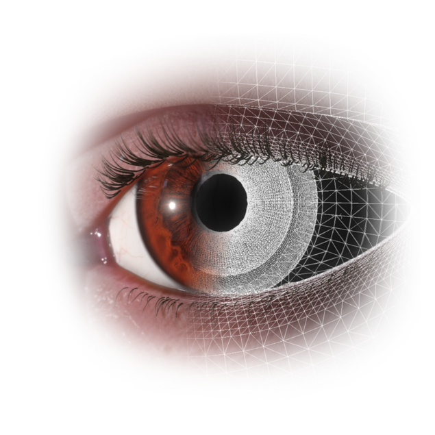
Physical models developed by Haag-Streit permit simulation of tissue properties and complex interactions between tissue, fluids, and surgical instruments in real time. Simulated behavior includes deformation, tearing, cracking, fluid dynamics, and subtle visual effects (e. g. red reflex in cataract surgery, distortions in retina surgery). The advanced algorithms allow for fast computation and almost latency-free results.

Binocular microscope

Stereo visualization

While training on Eyesi Surgical, users see the surgical field through a binocular microscope featuring mechanical and optical systems from Haag-Streit. The microscope offers a precise stereo visualization of the surgery simulation. Focus and zoom are operated using the microscope foot pedal.

Surgery instruments

True-to-life handpieces

The simulator’s handpieces are inserted through incisions in the model eye. The cataract patient head can be operated on from a temporal or superior position. Virtual cataract instruments, such as forceps, visco cannula, cystotome, and phaco probe can be assigned to the handpieces using the touch screen. For posterior segment surgery training, instruments such as a light probe, forceps, endolaser, or vitrector are available. In the retinal detachment training module, trainees can select an air, oil, or gas infusion.
OR machine

Interface and foot pedal

Eyesi Surgical features all common OR machine functions, including a venturi and peristaltic pump, and a two-axis foot pedal to control fluidics. Foot pedal settings can be configured according to individual preferences or to presets. Trainees can change the phaco or vitrectomy machine settings to explore the effects of parameter changes. The complex interaction between instruments and intraocular tissue can be experienced in real time.

Wide-angle viewing system

Auxiliary optics

To enhance the lifelike training environment of Eyesi Surgical for posterior segment surgery, it is possible to integrate a binocular indirect ophthalmomicroscope and a stereoscopic diagonal inverter hardware mimic, which is operated like a real OCULUS BIOM®/SDI®. The complex interactions of auxiliary optics are reproduced accurately in the simulation. Even an accidental contact of the lens with the cornea is reproduced.