Guidance & feedback

Designed for self-guided learning

Visual and auditory clues

Immediate feedback

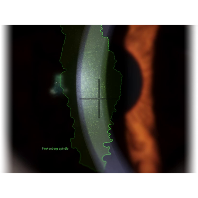
Eyesi Slit Lamp features visual and auditive guidance to support beginners in their learning process, for example, by highlighting anatomical or pathological findings or if crucial areas are not in focus.

Settings icons

Correct device handling

The slit lamp is a complex tool with many degrees of freedom. To foster correct device handling, Eyesi Slit Lamp provides users with an overview of correct and incorrect slit lamp settings to achieve a given examination target. Only if all slit lamp settings are correct is the pathological finding highlighted in the microscope, and a tile with medical background information appears on the instuctions screen.

Findings tiles

Recap of medical background information

Each time a trainee detects a pathological finding, it is highlighted, and a findings tile appears on the touch screen providing medical background information. All detected findings are stored in the trainee’s personal findings library for later recap of the learning matter.

In-microscope guidance

Fluid learning experience

The in-microscope view shows guidance elements depending on the current task. The instruction icons also displayed on the touchscreen are always visible in the microscope. The clearly recognizable overlay shows users all required slit lamp settings, allowing students to explore and adjust their settings without having to switch from eyepiece to screen, adding convenience and comfort to learning. In fundoscopy and gonioscopy tasks, a retina map and a 360-degree chamber angle overview provide orientation and highlight areas that still need to be examined.